Retina

Retina

Age-related Macular Degeneration

AMD is a common eye problem and is one of the primary causes of vision loss in people who are aged over 50 years. It damages the macula, which is a tiny area near the center of the retina. It is needed by the eye clear, central vision, to see objects that are straight ahead. Treatments such as injections, photodynamic therapy or laser surgery might be prescribed.


Diabetic Macular Edema (dme)

Diabetic Macular Edema (DME) is the condition where of fluid in the macula —part of retina that is responsible for detailed vision abilities— collects excessively due to leaks in blood vessels. It is usually treated with laser treatments.


Diabetic Retinopathy

People suffering from diabetes can have diabetic retinopathy. This condition occurs when high blood sugar levels cause damage to retinal blood vessels.The condition can lead to loss in vision. Treatment can include control of blood sugar and pressure through lifestyle and diet changes, specialized injections, laser surgery, or vitrectomy.


Floaters

Eye floaters are usually caused by age-related changes that occur as the vitreous substance inside your eyes becomes more fluid. Contact an eye specialist immediately if you notice a significant increase in eye floaters than usual, sudden presence of new floaters, flashes of light in the eye that has floaters, and darkness on the sides of your vision, as this may indicate a vision threatening condition.


Hypertensive Retinopathy

When a person’s blood pressure is too high, the walls of the retinal blood vessel can thicken, restricting blood flow. Over time, this can damage the retina’s blood vessels, restrict the retina’s functions. This is known as hypertensive retinopathy. The condition is controlled through lifestyle changes and medications.


Retinal Vein Occlusion

When a vein in the retina is blocked, it is unable to drain blood from the area. This leads to bleeding and leakage of fluid from the blocked blood vessels. This condition is retinal vein occlusion.Some of the treatments for retinal vein occlusion include VEGF drugs, intravitreal injections, focal laser therapy, or pan-retinal photocoagulation therapy.


Macular Hole

A macular hole is a minuscule hole in the macula region of the retina. It provides the clear, central vision that is required for activities like reading, driving, etc. Severe cases may require surgery for treatment.


Epiretinal Membrane/Macular Pucker

In this condition scar tissue forms on the eye’s macula, located in the center of the retina. A epiretinal membrane can cause blurred and distorted central vision. When the condition affects day to day functioning vitrectomy surgery may be recommended.


Macular Edema

This is the collection of fluid in the macula, of the retina. The liquid buildup causes distortion in the vision. Processes such as focal laser photocoagulation and drugs injected directly into the eye can be used for treatment.


Retinal Detachment

When the retina detaches, it is moved from its normal position. If not promptly treated, the condition can lead to permanent loss of vision laser surgery, vitrectomy or freeze treatments are commonly used for treatment.


Retinal Tear / Retinal Hole

Retinal holes and tears are tiny breaks in the retina that can show symptoms like cloudy, or blurry vision, or dark shadow in sides of vision. Laser photocoagulation, cryopexy, and diathermy are some processes that are commonly used to treat retinal tears.


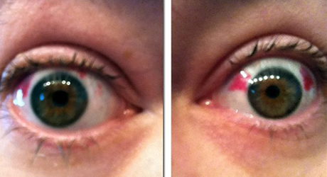
Retinal Hemorrhage

This is a disorder of the eye in which bleeding occurs in the tissue on the back of the eye. It can be caused due to hypertension, retinal vein occlusion or diabetes. If required laser surgery or anti-vascular endothelial growth factor (VEGF) drugs might be recommended.
Squint (Strabismus)

Squint is deviation of either eye from the midline position. Squint is not only cosmetically unappealing nut also impairs binocular single vision. Squint should be corrected as early as possible so that binocular single vision (BSV) may develop normally.
BSV is needed for distance and depth perception, for everyday activities like drawing, playing ball game and performing fine tasks. Squint can be covered easily with spectacles, exercises/or surgery. The child/person with squint many need to wear glasses and keep one eye occluded for a few months for exercise can be given on machine called synoptophore. Squint surgery is safe and does not involve opening up the eyeball like cataract (It is thus an extra ocular and not intraocular surgery). The patient needs to take eye drops for a month after surgery.

  • Exotropia- outward deviation of eye.
  • Esotropia- inward deviation of eye.

Glaucoma

Glaucoma (Kachbindu in Marathi, kalamoti in Hindi) involves increase in the intraocular pressure IOP (eyes pressure) Normal IOP is 12-19 mm of hg, if the IOP is raised more than 20 mm of hg. It may put in the optic nerve which convey the single from the eye to the ——.
This loss of nerve function could lead to irreversible vision loss side vision is affected first and than central vision. As few decades ago glaucoma was associated with blindness but with modern diagnostic and therapeutic techniques, it can be easily controlled. The patient needs to get his eyes checked up regularly with measurement of IOL once every Six month visual fields (side vision) needs to be checked with a auto perimeter. This test takes up to half an hour.
Instruments needed
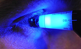
Application tonometer to measure IOP slit lamp with gonioscopy – to see angle of anterior chamber, Autoperimetry – for visual fields testing Glaucoma is easily treated with medicine, laser and /— surgery (trabeculectomy).

Contact Lens

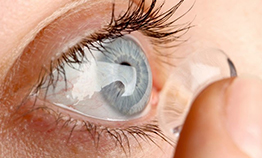
Contact lenses are a very convenient alternative to wearing spectacles. They enable visual freedom and full peripheral vision that is impossible to achieve with spectacles. They are ideal for sports and can work to help create a whole new look. Everyone has different visual needs, and as a result we fit different kinds of contact lenses. If you are new to contact lenses or regular wearer, we have the lenses for you: A good way to look at disposable lenses is by how often you would like to replace them:

Types of Contact Lenses : Soft Contact Lenses

New lenses are inserted every time the lenses are worn, and are thrown away at the end of the wearing day. Great for casual wear to play sport, attend special functions or wear on holiday.

Lenses are worn during waking hours, then removed and cleaned before sleep and replaced either fortnightly or monthly. Suitable to people who want to wear contact lenses every day for up to 10 hrs.

This type of lens is worn for longer working hours (> 10 hours) and replaced monthly or biweekly. They may suit people with busy lifestyles where these lenses are an ideal option.

Semisoft or rigid gas permeable contact lenses are also good alternative to soft contact lenses. Thinking Contact Lenses – Think Dr. Gogate’s eye clinic – For Expertise, Advice and Service offered by Dr Meena Kharat

Contact Lens Trial set:

  • RGP contact lens set
  • Soft contact lens set
  • Miniscleral lens trial set
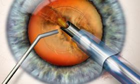
cataract-surgery-thumb

Uveitis and HIV

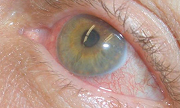
The clinic offers special facilities for HIV affected individuals. Uveitis is inflammation of uvea, the middle layer of the eye between the sclera and the retina. Most treatment may be long drawn.

LASIK Laser Eye Surgery

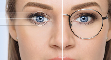
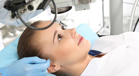
LASIK (LASER Assisted In-Situ Keratomileusis) is the most common laser procedure done on the eye to reduce and eliminate refractive error. More than 100,000 people a year now have this procedure performed in the UK.
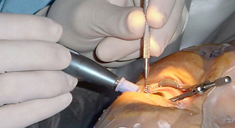
During LASIK, an eye surgeon cuts across the cornea of the eye and raises a flap of tissue and carries out reshaping to correct vision.
LASIK is often used for people who are short-sighted, long-sighted, or who have astigmatism. LASIK, may not be suitable for people with strong lens prescriptions, such as a high degree of short-sightedness.
No medical procedure is without risks, and complications can happen in fewer than 1 in 50 laser eye operations.
One in 3 people patients will still need to glasses some of the time  after the procedure, such as for night driving

  • It works and is proven to correct vision in most cases.
  • LASIK laser eye surgery is associated with almost no pain.
  • Vision  is corrected almost immediately or by the day after LASIK laser eye surgery.
  • Recovery is quick and usually no bandages or sutures are required after LASIK laser eye surgery.
  • Adjustments can be made years after LASIK laser eye surgery to further correct vision.
  • After having LASIK laser eye surgery, most patients no longer need corrective eyewear.

Despite the pluses, there are some disadvantages:

  • Changes made to the cornea cannot be reversed after LASIK laser eye surgery.
  • Corrections can only be made by additional LASIK laser eye surgeries.
  • LASIK laser eye surgery is technically complex. Problems may occur when the doctor cuts the flap, which can permanently affect vision.
  • LASIK laser eye surgery can cause a loss of “best” vision with or without glasses at one year after surgery. Your best vision is the highest degree of vision that you achieved while wearing your contacts or spectacles.

Some patients experience discomfort in the first 24 to 48 hours after surgery.
Other side effects, although rare, may include:

  • Glare
  • Seeing halos around images
  • Difficulty driving at night
  • Fluctuating vision
  • Dry eyes

Before your LASIK laser eye surgery, you will meet with an adviser who will discuss what you should expect during and after the surgery. During this session, your medical history will be evaluated and your eyes will be tested. Likely tests include measuring corneal thickness, refraction, and pupil dilation. At any meeting with your surgeon, you should ask further questions you may have. Afterwards, you can book an appointment for the procedure.

IPCL SURGERY

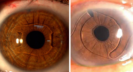
Implantable Phakic intraocular lenses, or phakic lenses, are lenses made of plastic or silicone that are implanted into the eye permanently to reduce a person’s need for glasses or contact IPCL SURGERY lenses. Phakic refers to the fact that the lens is implanted into the eye without removing the eye’s natural lens. During phakic lens implantation surgery, a small incision is made in the front of the eye. The phakic lens is inserted through the incision and placed just in front of or just behind the iris.

Phakic lenses are used to correct refractive errors, errors in the eye’s focusing power. All phakic lenses approved by the FDA are for the correction of nearsightedness (myopia). The cornea and natural lens of the eye focus light to create an image on the retina, much like the way the lens of a camera focuses light to create an image on film. The bending and focusing of light is also known as refraction. Imperfections in the focusing power of the eye, called refractive errors, cause images on the retina to be out of focus or blurred.

People who are nearsighted have more difficulty seeing distant objects than near objects. For these people, the images of distant objects come to focus in front of the retina instead of on the retina.

Ideally, phakic lenses cause light entering the eye to be focused on the retina providing clear distance vision without the aid of glasses or contact lenses.

Surgery is not required to correct nearsightedness. You can wear glasses or contact lenses instead to correct your vision. Depending on how nearsighted you are, and other conditions of your eye, other refractive surgery (surgery to correct refractive errors) options may be available to you, including PRK (Photorefractive Keratectomy) and LASIK (Laser Assisted In-Situ Keratomileusis).

Phakic lenses are intended to be permanent. While the lenses can be surgically removed, return to your previous level of vision or condition of your eye cannot be guaranteed.

Phakic intraocular lenses are implanted in the eye without removing the natural lens. This is in contrast to intraocular lenses that are implanted into eyes after the eye’s cloudy natural lens (cataract) has been removed during cataract surgery.

The first part of the anatomy of the eye animated illustration shows an external side view of the eyeball with the parts labeled. The sclera, cornea, iris, pupil, and optic nerve are identified. The second part of the illustration shows an internal side view with the eye cut in half from front to back. It identifies the sclera, cornea, anterior chamber, iris, pupil, posterior chamber, natural lens, retina, and optic nerve.

The illustration of how phakic lenses work shows the side view of eye as if you cut it in half from front to back and you were looking at the inside. It shows light rays from a stop sign being focused by the cornea and lens onto the retina of an eye that needs no vision correction. It depicts the clear retinal image of the stop sign being sent to the brain by the optic nerve. When light rays come into focus on the retina, we see the object clearly.

The image of the same stop sign is focused in front of the retina in a nearsighted (myopic) eye. The light rays from the stop sign that fall onto the retina are out of focus creating a blurred image. The blurred retinal image is sent to the brain by the optic nerve. When light rays do not come into focus on the retina, we see the object as blurred.

The inset image shows the clear retinal image of the stop sign being sent to the brain by the optic nerve. This is the idealized results of the phakic lens bending light from the stop sign to redirect the focused image onto the retina. When light rays are redirected by the phakic lens onto the retina, the nearsighted eye sees distant objects more clearly.